Vesicovaginal fistula
Applied Radiology — Vol. 37 , Issue 5 , pp. 42 -44
DOI: 10.37549/AR1608
Published: May 1, 2008
Categories
CASE SUMMARY
A 44-year-old woman with a suspicious uterine mass underwent an abdominal hysterectomy. She experienced vaginal discharge and recurrent lower urinary tract infections for 2 weeks after the operation. At a 2-year follow-up and evaluation of this patient, all imaging procedures (including the instillation of methylene blue solution, conventional ultrasound, cystoscopy, and intravenous pyelogram [IVP]) were normal. Subsequently, the patient was referred to our nuclear medicine ward for a radionuclide cystogram.
IMAGING FINDINGS
The results of the radionuclide cystogram are illustrated in Figure 1. In addition, by measuring the accumulation within the tampon that had been placed in the upper vaginal portion before beginning the study, abnormal radiotracer accumulation in this region was confirmed. In the filling phase, an abnormal focus of radiotracer activity was observed in the posteroinferior aspect of the bladder, which was compatible with the region of the upper vagina. The focus of abnormal radiotracer activity was clearly visualized in the lateral view (Figure 1).
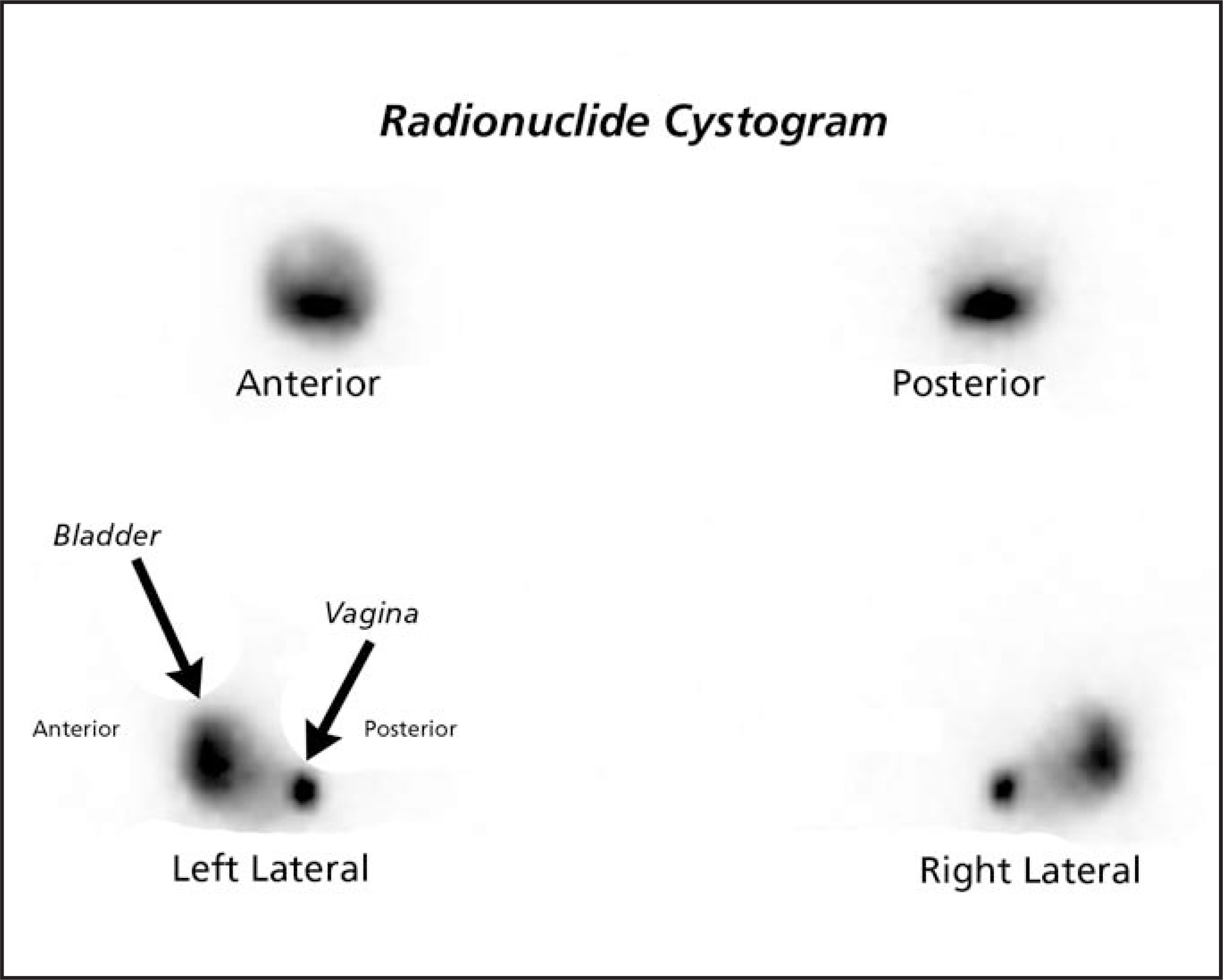
On the basis of the scintigraphic findings, the most likely diagnosis was vesicovaginal communication. The other probable diagnosis was downward posterior-located diverticulum of the bladder, but this was ruled out by the quantitatively measured vaginal tampon. The diagnosis of vesicovaginal communication (fistula) was quantitatively confirmed by the proportion of tampon activity before and after the technetium (Tc)-pertechnetate instillation to the bladder.
DIAGNOSIS
Vesicovaginal fistula
DISCUSSION
Vesicovaginal fistulas are among the most common types of fistulas in uro gynecology.1 In developed nations, these fistulas are usually unfortunate complications of gynecologic or other pelvic surgeries and radiotherapy.2 In contrast, in underdeveloped countries, birth trauma is the major cause.2 There are only a few reports of fistulas developing spontaneously without any previous intervention or trauma.3
The following diagnostic tools are established to evaluate vesicovaginal fistulas: pyridium tablets, the instillation of methylene blue solution, cystogram, vaginoscopy, urethrocystoscopy, vaginogram,3 and even computed tomography.4 However, all of these methods have their own limitations and are not completely reliable for the detection of these path ologies. Conventional ultrasound is also of no help in diagnosing vesicovaginal fistulas, but some success has been reported using color Doppler ultrasound and ultrasound contrast media.3
CONCLUSION
To the best of our knowledge, this is the first report of a case of vesicovaginal fistula that was diagnosed with radionuclide cystography. With this finding, we suggest that as an alternative approach to other currently used diagnostic approaches, it is possible to detect these pathologies with the use of radio nuclide cystography. As it is a common drawback in all of nuclear medicine procedures, the lack of spatial resolution of this method limits its ability to localize the exact anatomical site of the fistula, which is an important issue in surgical management of the disease.1 In cases in which the presence of a fistula is suspected and other imaging modalities cannot confirm or exclude the development of a fistula, a radionuclide cysto gram could be useful. This approach must be addressed in larger series of patients, and correlation with other imaging modalities is needed. A prospective study is currently being performed to confirm this approach and these data.
References
- Angioli R, Penalver M, Muzii L. Guidelines of how to manage vesicovaginal fistula. Crit Rev Oncol Hematol. 2003;48:295-304.
- Cortesse A, Colau A. Vesicovaginal fistula [in French]. Ann Urol (Paris). 2004;38:52-66.
- Volkmer B, Kuefer R, Nesslauer T. Colour Doppler ultrasound in vesicovaginal fistulas. Ultrasound Med Biol. 2000;26:771-775.
- Peters J, Lörcher U. Computerized tomography of a vesico-vaginal fistula caused by a foreign body [in German]. Zentralbl Gynakol. 1991;113:789-793.
Citation
. Vesicovaginal fistula. Applied Radiology. 2008;37(5):42-44. doi:10.37549/AR1608.