Virtual bronchoscopy: Technical features and clinical applications
Applied Radiology — Vol. 32 , Issue 4 , pp. 33 -39
DOI: 10.37549/AR1180
Published: April 1, 2003
Categories
In the clinical evaluation of pulmonary disease, fiberoptic bronchosopy is a crucial tool in the diagnosis of a variety of chest diseases. Though often instrumental in the diagnosis of a variety of neoplastic, inflammatory, and infectious diseases, fiberoptic bronchscopy (FOB) can have important limitations: it is invasive and time-consuming, and it requires sedation.1 It may not be tolerated in the young, in the critically ill, or in patients with coagulopathies. In patients with significant airway disease/stenoses, bronchoscopic evaluation of the airway distal to areas of stenoses/narrowing is technically difficult and may compromise patient oxygenation significantly. Equally important, the evaluation of extraluminal pathology is significantly limited in fiberoptic bronchoscopy.2
The last decade has seen incredible advances in thoracic imaging. The advent of spiral computed tomography (CT) and the acquisition of volumetric data sets have allowed anatomic depiction of axially acquired data. Multiplanar reformatting (MPR), maximum intensity projection (MIP), and volume-rendering techniques are now standard in chest imaging. With increasingly sophisticated software, axial images can be reconfigured to display this data from an endoscopic perspective. This has clearly been important in the evaluation of bowel pathology, and virtual colonoscopy has provided impressive sensitivity in the noninvasive screening evaluation of colon cancer. Virtual bronchoscopy (VB) has similarly become an important tool in the evaluation of chest diseases. This presentation will outline these advances and will present the role of VB in the evaluation of a variety of neoplastic and non-neoplastic processes.
While original virtual endoscopy programs were often time-consuming and impractical to the practicing radiologist, increasingly sophisticated postprocessing techniques have improved the speed and accessibility of the user interface, enabling rapid virtual endoscopic depiction of the airways, pulmonary arteries, and aorta. These expanded imaging capabilities combine to enhance our ability to display complex thoracic pathology, serve our referring clinicians, and enhance patient care. Several principles in image acquisition are crucial in the successful performance of virtual bronchosopy. A number of studies have demonstrated the importance of narrow collimation and reconstruction overlap of at least 50% to optimize any type of three-dimensional imaging3; and these parameters are similarly important to virtual bronchoscopic evaluation. For single-slice CT scanners, a slice thickness of 3 to 5 mm is preferred. While pitch values up to 2 are usually acceptable for the volumetric depiction of pathology, most published protocols prefer a pitch of 1 for virtual bronchoscopic rendering using a single-slice scanner. With the advent of multislice scanning, improved and gantry rotation times of 500 msec, virtual bronchoscopic imaging can now be routinely performed with 1-mm slice collimation.
While the use of intravenous (IV) contrast is not necessary in most cases performed for VB, it can be very helpful in the depiction of extraluminal pathology. To optimize contrast opacification of extraluminal vascular structures, 100 to 140 mL of IV contrast is used at injection rates that range from 2 to 5 mL/sec.
As will be discussed later, accurate integration of extraluminal pathology with the virtual bronchscopic data helps optimize VB diagnostic capabilities. Accurate segmentation of extraluminal structures is optimized with IV contrast and is therefore important to advanced VB protocols. At our institution, patients for whom VB is considered are now scanned entirely on our multislice scanners (Philips MX8000, Philips Medical Systems, Best, Netherlands). Decisions on 1- or 2.5-mm scanning depend on the clinical question, anatomic coverage required, and radiation dose to the patient. The speed of multidetector technology allows dynamic inspiratory/ expiratory endoscopic imaging, which has proven important to our referring pulmonologists in the evaluation of tracheomalacia and suspected upper-airway collapse.4
We have recently acquired a 16-slice CT scanner (MX IDT, Philips Medical Systems). This has allowed virtual bronchoscopic evaluations using 1-mm slice thickness, often with imaging times <4 seconds. This rapid imaging allows outstanding endoscopic renderings in the most critically ill patient.
Future research in image optimization in VB includes the role of cardiac gating in the accurate depiction of airway stenosis. Following the acquisiton of the spiral CT data, transfer of the DICOM data to advanced imaging workstations is necessary. There is a variety of advanced imaging products capable of virtual endoscopic imaging. These products include General Electric Navigator (GE Medical Systems, Milwaukee, WI), Vital Images Voxel View (Vital Images, Fairfield, CT), Iris Explorer (Silicon Graphics, Mountain View, CA).5 A number of advanced, hybrid imaging techniques have been developed at individual institutions to further optimize the VB data. At our institution, we have used Voyager Software (Phillips Medical Systems) for VB. The recent transition to a Windows NT workstation has significantly improved the speed for image reconstruction of this data and has made virtual bronchoscopic imaging easily accessible even to the least experienced operator.
The effective use of VB necessitates an understanding of the anatomy seen during fiberoptic bronchoscopy. In particular, the perspective of the bronchoscopist is opposite the traditional orientation of the radiologist: the bronchoscope displays airway anatomy in a cranial-caudal direction, with the patient in a supine position (Figure 1). For radiologists involved with VB, a solid understanding of this anatomic perspective is important, and active participation/correlation with fiberoptic bronchoscopy is helpful in understanding the capabilities and limitations of VB. In most VB software packages, both surface and volume rendering can be performed. Surface rendering has been used routinely because reconstruction times are considerately faster than volume rendering techniques. Standard threshold values for surface-rendering techniques have been described.6 While these thresholds are effective in the main and lobar bronchi, artifacts can occur in the smaller airways, and may also result in an overestimation of stenosis.7 Currently, faster computer reconstruction times and more flexible thresholding capabilities have made volume-rendered VB our technique of choice in the evaluation of the airway.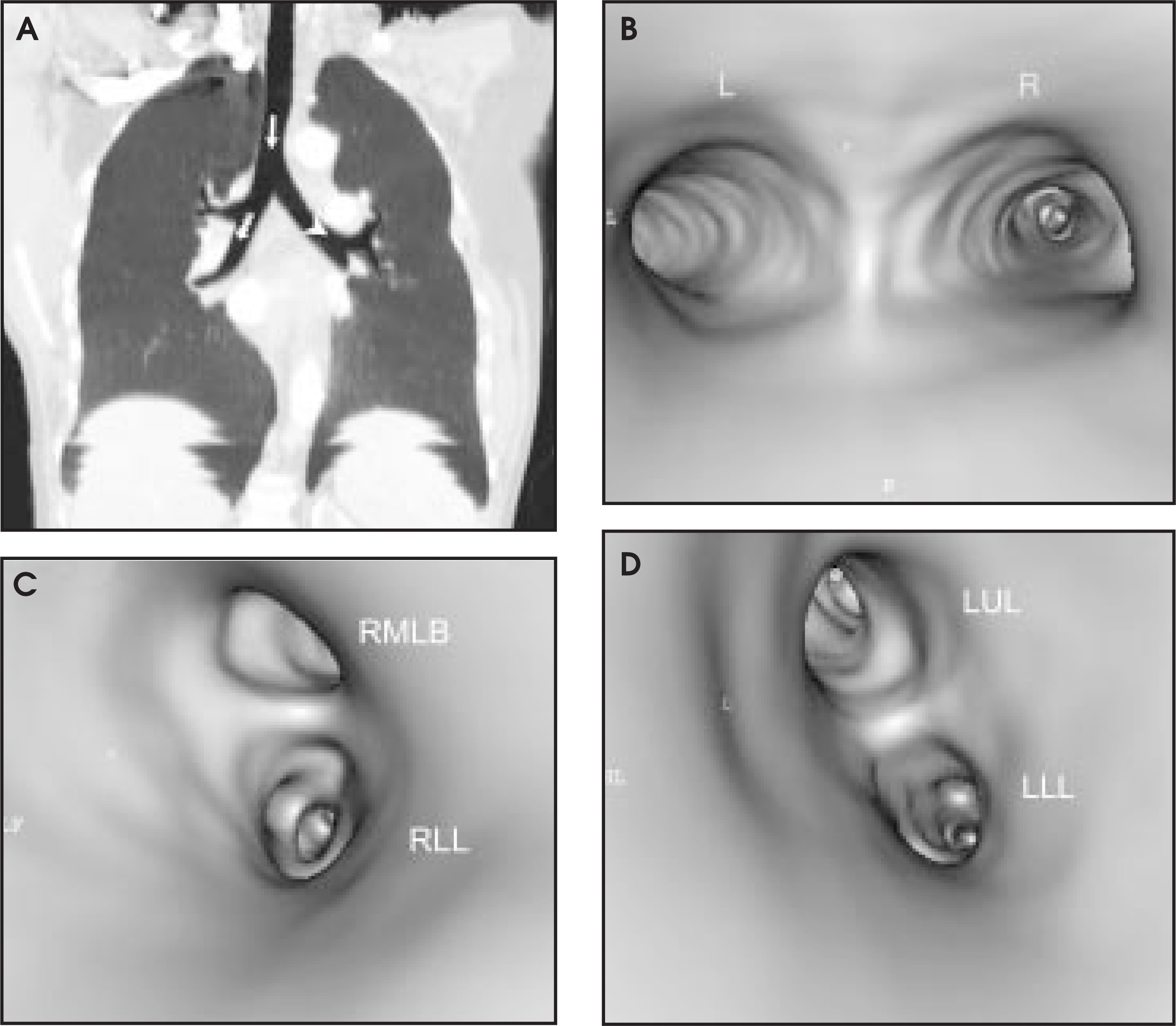
A number of papers have been written comparing the diagnostic value of VB with standard CT images, as well as with fiberoptic bronchoscopy. One of the values of virtual endoscopy in the airways is the evaluation of the upper airway (Figure 2). In a study of 30 patients, Burke et al8 evaluated the use of VB for the assessment of airway obstruction. This group included patients with upper airway disease ranging from airway stenosis, laryngo tracheomalacia, tumors, and webs (Figure 3). There was excellent agreement between VB and FOB in the evaluation of airway stenoses.8 The differences in ratios of stenoses/lumen were within 10% for VB versus upper airway endoscopy. The evaluation of dynamic airway collapse was much less reliable with virtual endoscopy, but there was a clear role for virtual endoscopy in airway stenoses.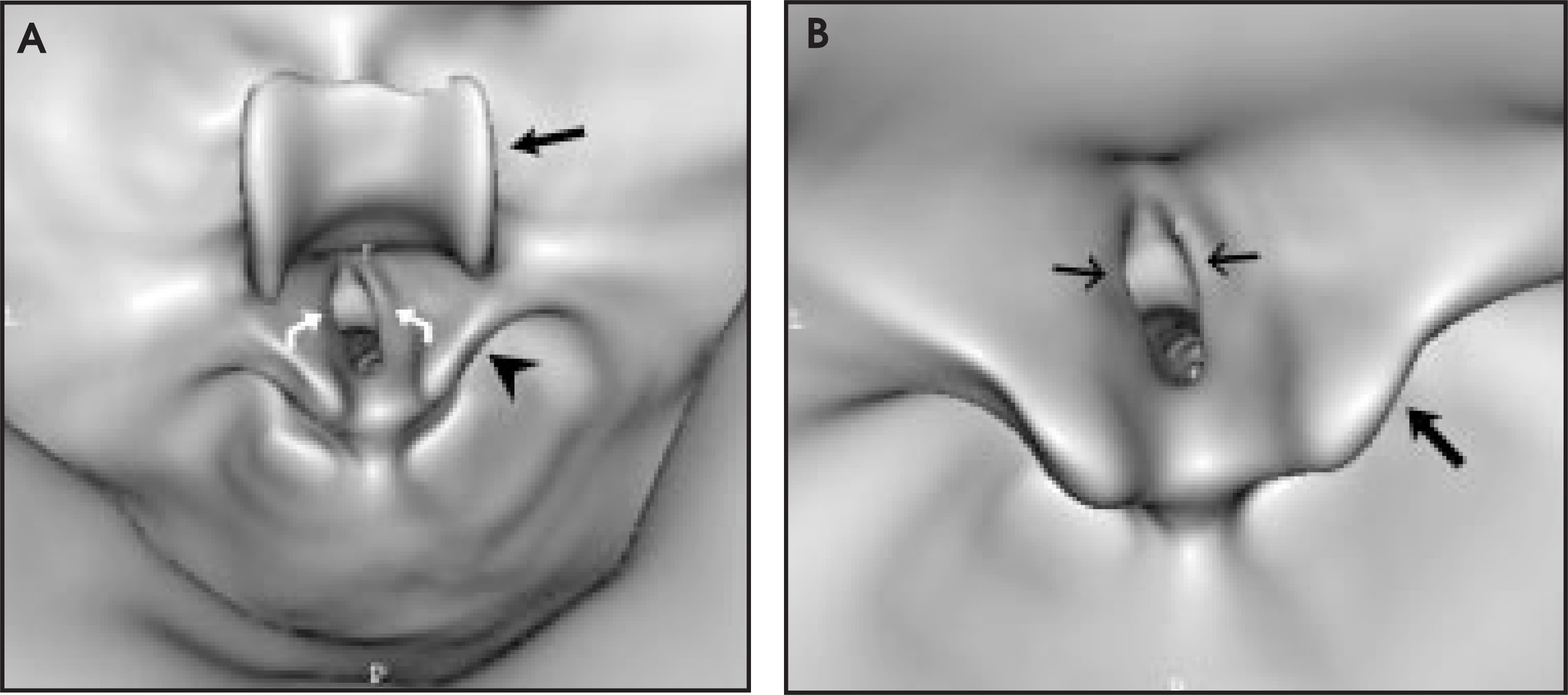
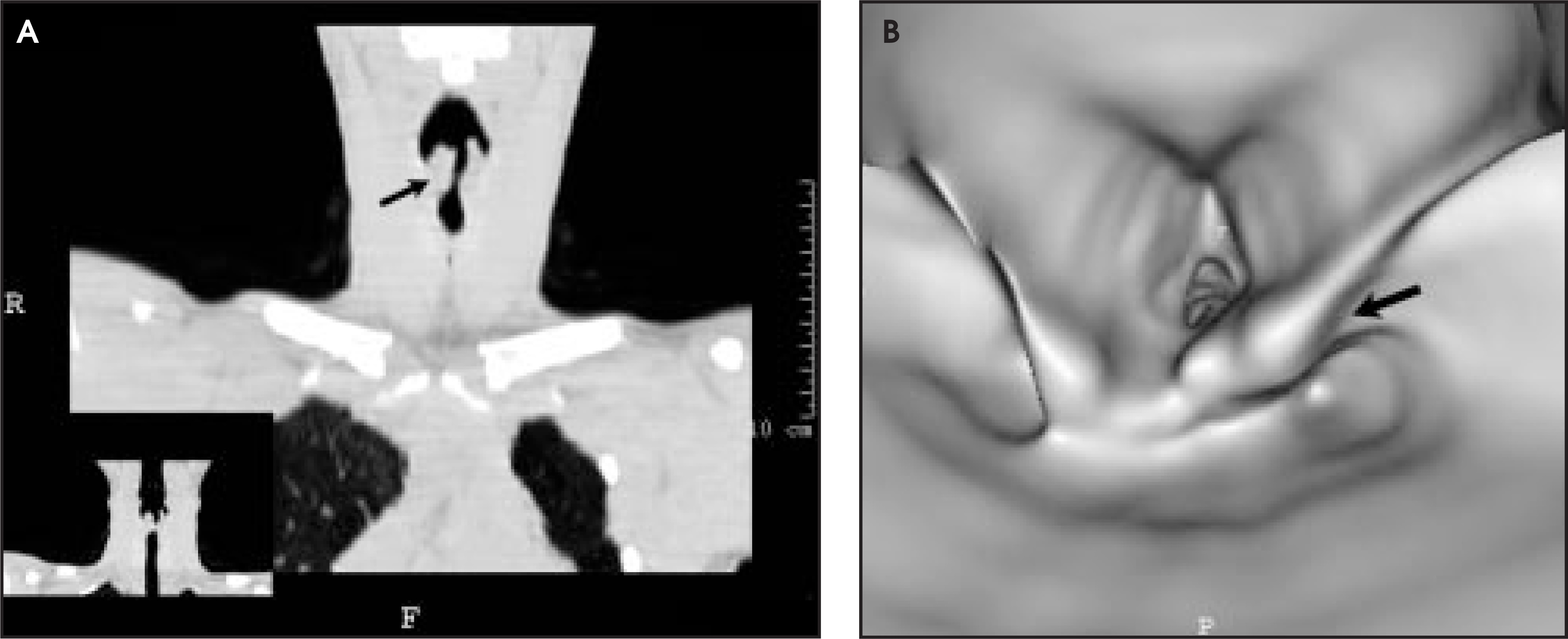
Several papers have been written defining the role of VB in patients with airway stenoses secondary to bronchogenic carcinoma. Liewald et al9 reported the results of their study of 30 patients with bronchogenic carcinoma. While central lesions were well identified by VB, stenosis secondary to smaller lobar lesions were not identified accurately. Rapp-Bernhardt and colleagues10 evaluated a number of visualization techniques, including axial, MPR, minimum intensity projection (MinIP), and VB. Virtual bronchoscopy evaluation of stenoses showed the lowest interobserver variability. While there was no significant difference between virtual and fiberoptic bronchoscopy in the estimation of stenoses, VB tended to overestimate the degree of stenosis.10 This was believed to be due to thresholding limitations of stenoses in VB. In addition, mucosal abnormalities were not evaluated well with VB.
With newer 3D segmentation techniques, the visualization of this endobronchial and mucosal pathology has been enhanced. These advanced techniques have become important in the surgical planning of patients with complex neoplastic involvement of the airway (Figure 4). There are increasing reports in the literature on the use of VB in improving the diagnostic accuracy of fiberoptic bronchoscopy. In a paper by McAdams et al,11 VB was reviewed by the pulmonologist for evaluation of lymph node biopsy, and was preferred to the axial CT slices. There was a slight improvement in diagnostic yield compared with standard axial images when VB images were reviewed.11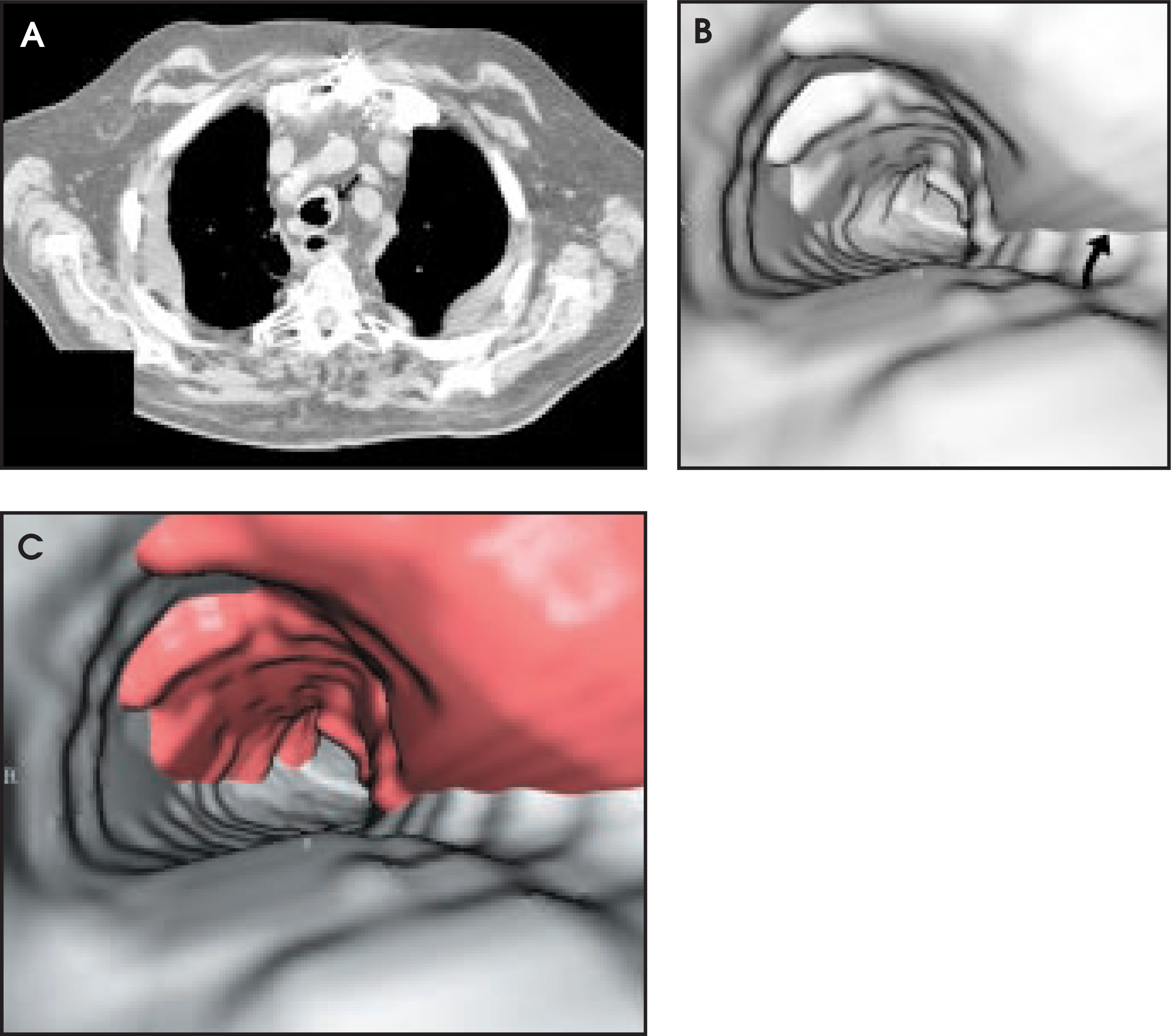
Hopper and coworkers12 have described a technique of lymph-node highlighting. Mediastinal lymph nodes were segmented manually, highlighted, and integrated into the surface-rendered VB.12 By varying the transparency of the bronchial wall, the involved lymph node could be chosen before the procedure. While there was no significant difference in the diagnostic sampling of subcarinal lymph nodes, these techniques improved diagnostic yield significantly for hilar and pretracheal lymph nodes. We have used this technique successfully in the preoperative evaluation of extraluminal tumor involvement (Figure 5).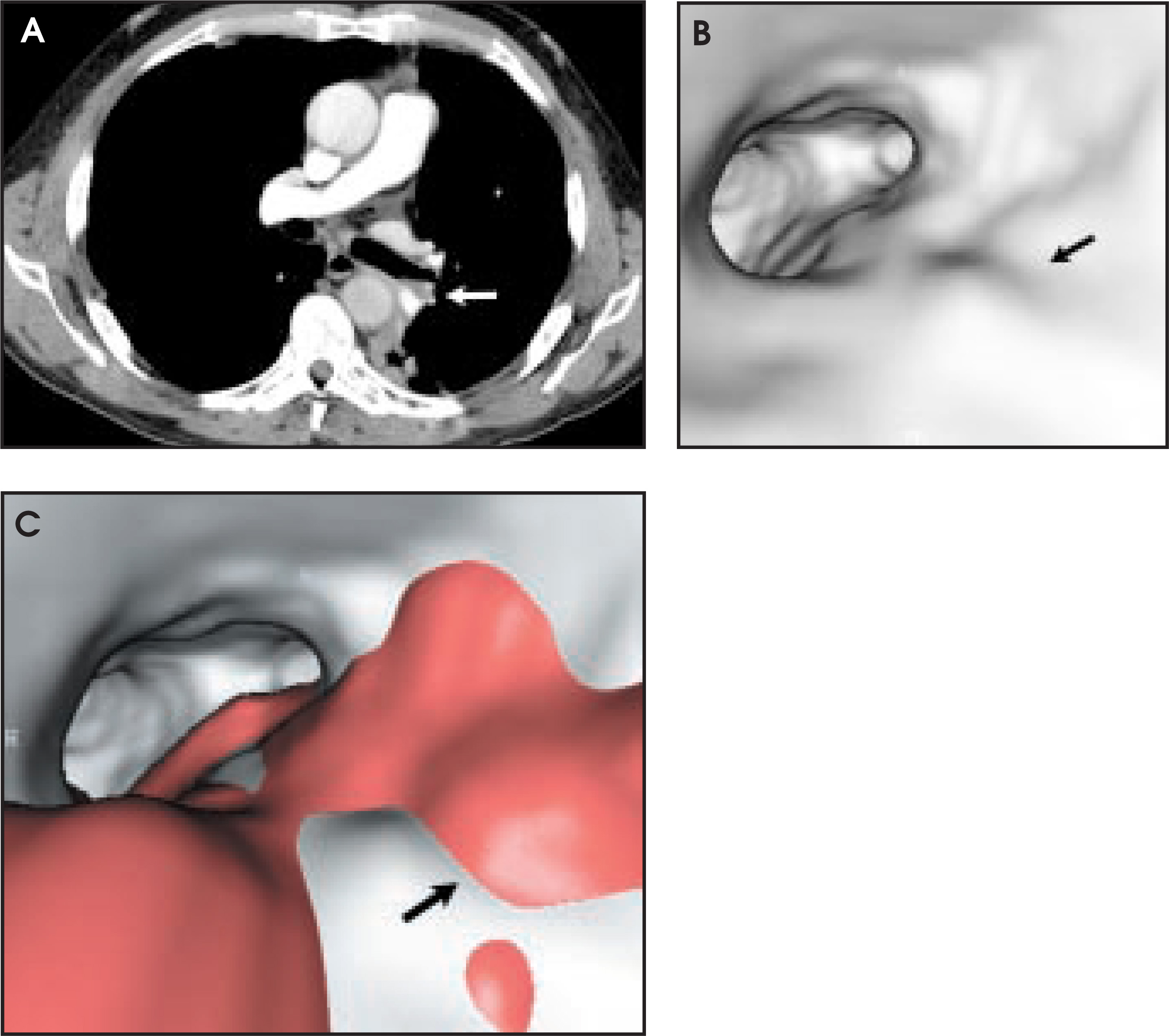
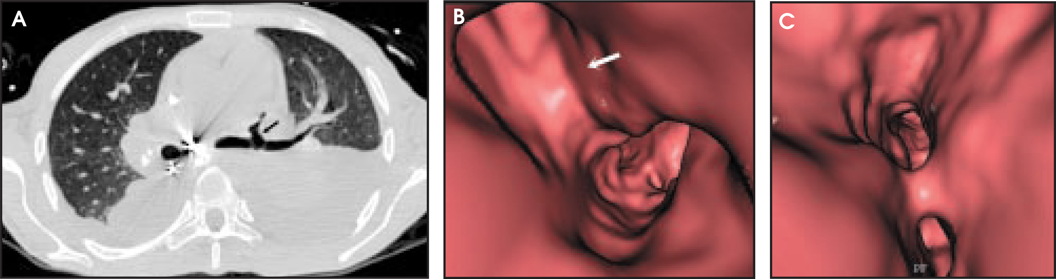
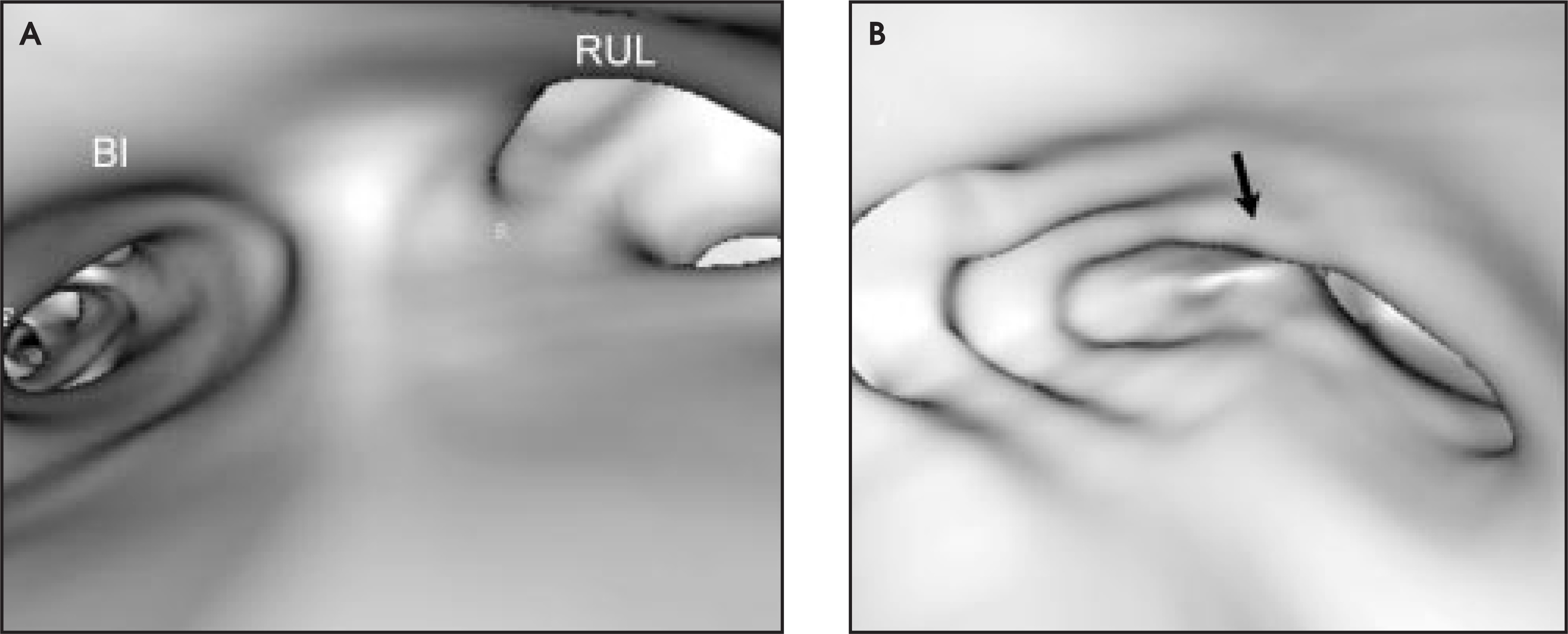
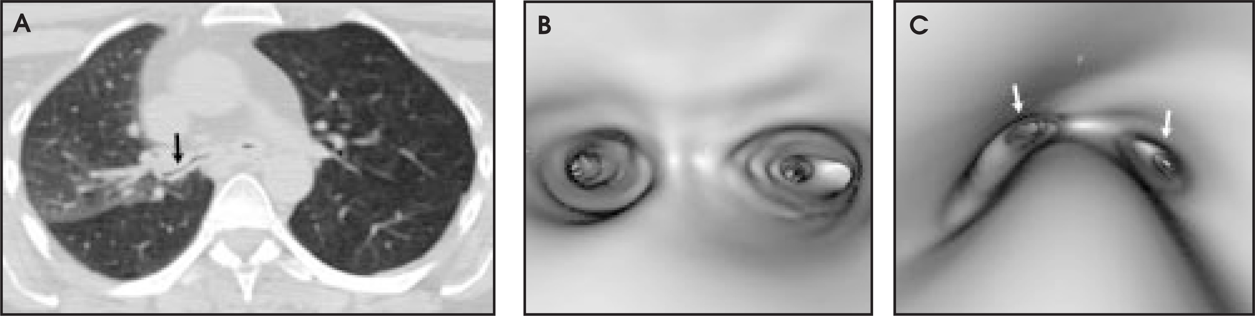
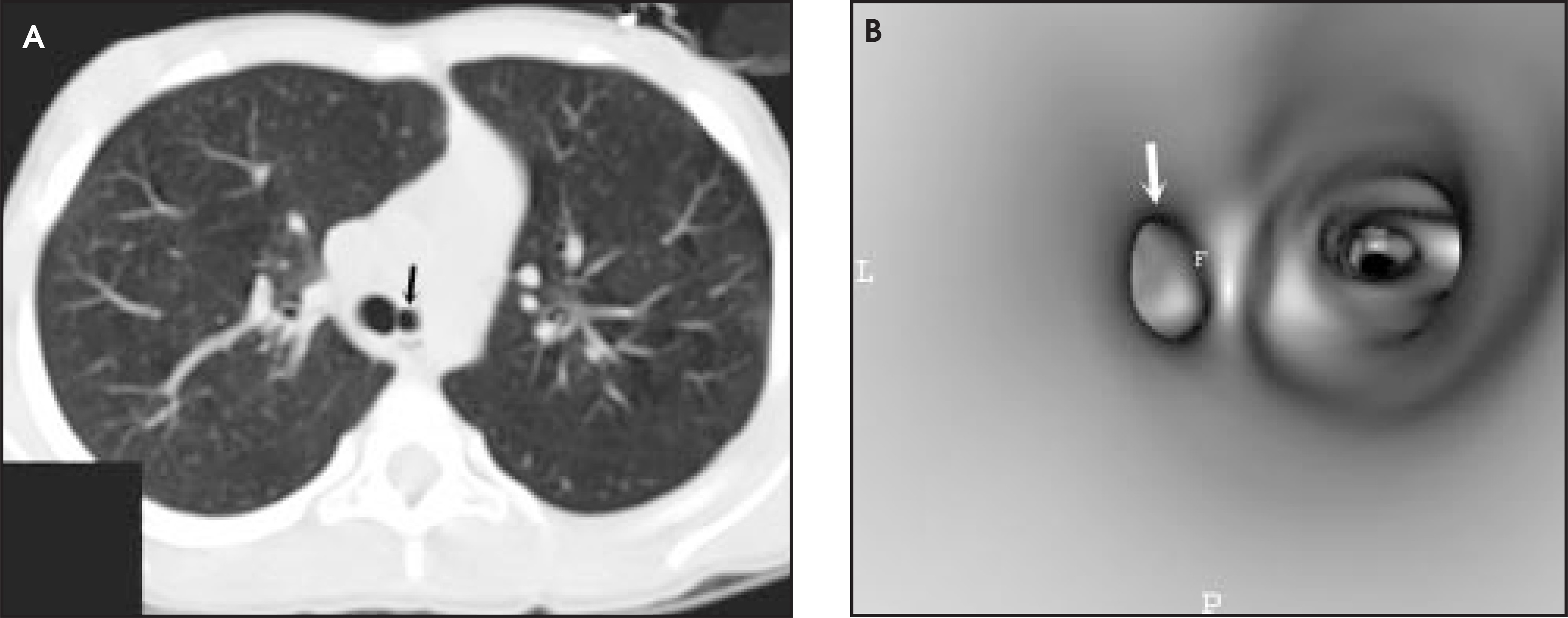
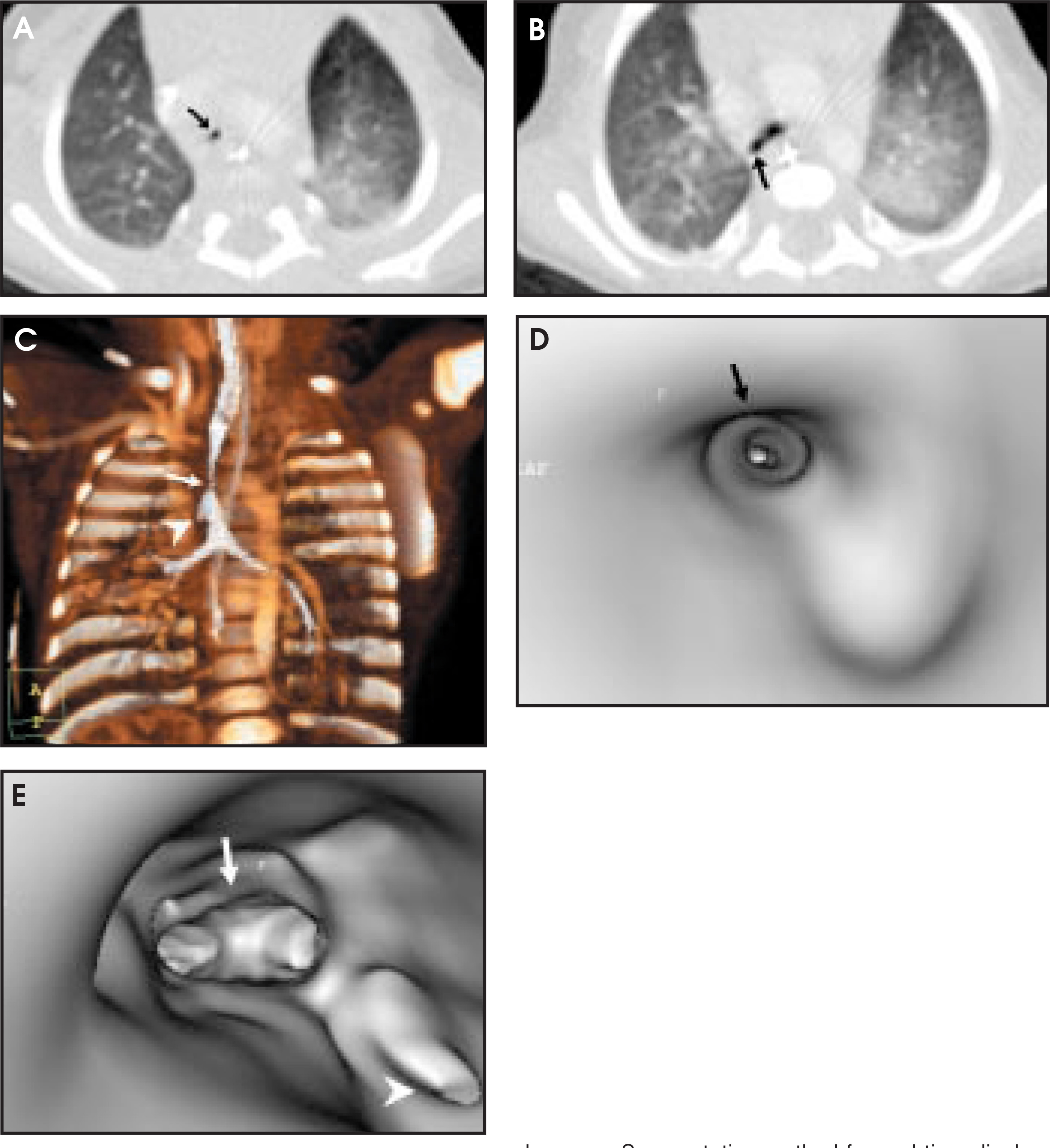
The evaluation of non-neoplastic airway disease has become an important source of physician referrals at our institution. Summers et al13 have written about the role of VB in the evaluation of patients with Wegener’s granulomatosis. The complex, multifocal nature of this disease is well suited for evaluation by VB, especially in patients with multifocal involvement (Figure 6). McAdams and colleagues14 have studied the role of VB in patients following lung transplantation. Stenoses at the anastomotic sites were better visualized with VB than with axial CT, MinIP, or MPR techniques. Determination of the degree of airway stenosis by VB resulted in the lowest interobserver variability of any of the techniques studied. Other complications of lung transplantation have become important indications for VB in our transplant population, including anastomotic dehiscence (Figure 7) and tracheobronchomalacia (Figure 8). Dynamic airway collapse is not limited to the lung transplant population and has become a major cause of referrals at our institution for the evaluation of tracheomalacia (Figure 9). One of the most important indications for VB at our institution is the evaluation of the problematic pediatric patient (Figure 10).15 With multidetector technology and close attention to radiation dose, the most complicated pediatric airway can be well imaged by VB (Figure 11).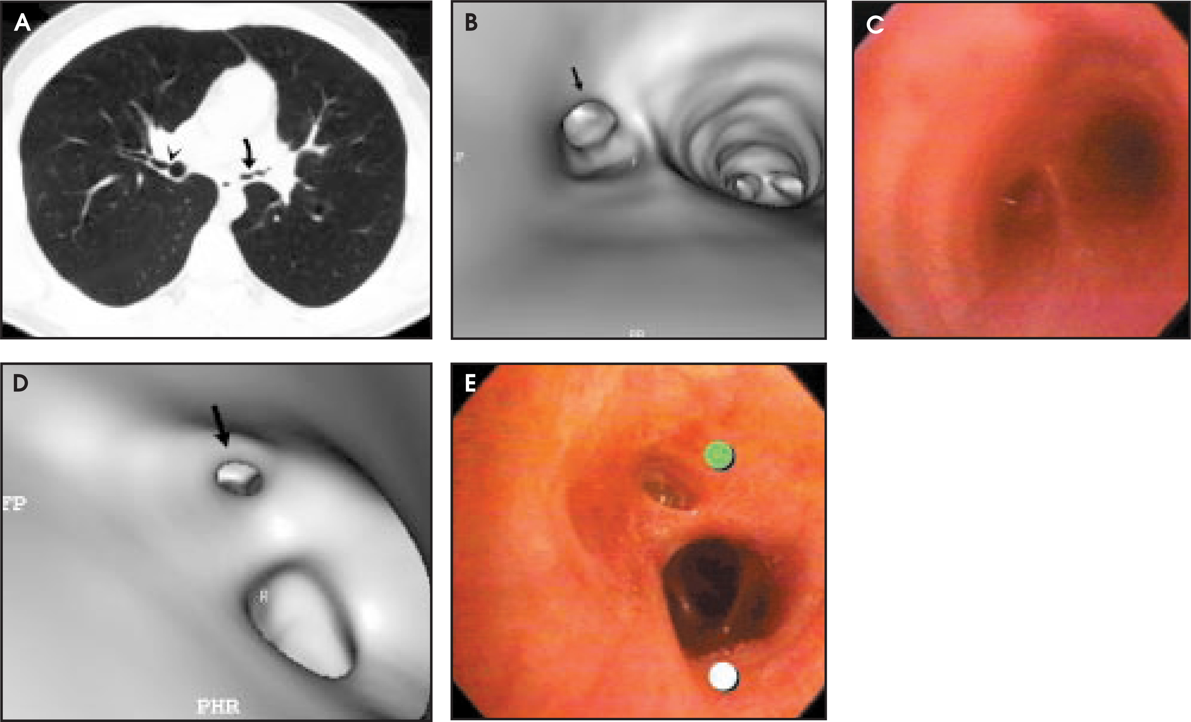
Conclusion
This paper has described the increasing capabilities of VB in the evaluation of patients with a variety of airway pathologies. As our imaging times decrease and 3D techniques become more sophisticated, the role of VB will continue to grow. An understanding of this technique will improve our understanding of pulmonary disease, and will serve to increase referring clinicians’ use of CT in the evaluation of complex airway disease. AR
References
- Pue C, Pacht E. Complications of fiberoptic bronchoscopy at a university hospital. Chest. 1995;107:430-432.
- Finkelstein S, Summers R, Nguyen D. Virtual bronchoscopy for evaluation of malignant tumors of the thorax. J Thorac Cardiovasc Surg. 2002;123:967-972.
- Ferretti G, Vining D, Knoplioch J, Coulomb M. Tracheobronchial tree: Three-dimensional spiral CT with bronchoscopic perspective. J Comput Assist Tomogr. 1996;20:777-781.
- Gilkeson R, Ciancibello L, Hegal R. Tracheobronchomalacia: Dynamic airway evaluation with multidector computed tomography. AJR Am J Roentgenol. 2000;176:205-210.
- Haponik E, Aquino S, Vining D. Virtual bronchoscopy. Clin Chest Med. 1999;20:201-217.
- Summers R, Shaw D, Shelhamer J. CT virtual bronchoscopy of simulated endobronchial lesions: Effect of scanning, reconstruction, and display setting and potential pitfalls. AJR Am J Roenthenol. 1998;170:947-950.
- Summers R, Feng D, Holland S. Virtual bronchoscopy: Segmentation method for real-time display. Radiology. 1996;200:857-862.
- Burke A, Vining D, McGuirt W. Evaluation of airway obstruction using virtual endoscopy. Laryngoscope. 2000;110:23-29.
- Liewald F, Lang G, Fleiter T. Comparison of virtual and fiberoptic bronchoscopy. J Thorac Cardiovasc Surg. 1998;46:361-364.
- Rapp-Barnhardt U, Welte T, Doehring W. Diagnostic potential of virtual bronchoscopy: Advantages in comparison with axial CT slices, MPR and MinIP. Eur Radiol. 2000;10:981-988.
- McAdams H, Goodman P, Kussin P. Virtual bronchoscopy for directing transbronchial needle aspiration of hilar and mediastinal lymph nodes: A pilot study. AJR Am J Roentgenol. 1998;170:1361-1364.
- Hopper K, Lucas T, Gleeson K. Transbronchial biopsy with virtual CT bronchoscopy and nodal highlighting. Radiology. 2001;221:531-536.
- Summers R, Aggarwal B, Sneller M. CT virtual bronchoscopy of the central airways in patients with Wegener’s granulomatosis. Chest. 2002;121:242-250.
- McAdams H, Palmer S, Erasmus J. Bronchial anastomotic complications in lung transplant recipients: Virtual bronchoscopy for noninvasive assessment. Radiology. 1998;209:689-695.
- Sorantin E, Geiger B, Lindbichler F. CT-based virtual tracheobronchoscopy in children—comparison with axial CT and multiplanar reconstruction: Preliminary results. Pediatr Radiol. 2002;32:8-15.
Citation
. Virtual bronchoscopy: Technical features and clinical applications. Applied Radiology. 2003;32(4):33-39. doi:10.37549/AR1180.