Virtual Rounds: Malignant Melanotic Nerve Sheath Tumor of the Neck 1
ARO Student Scan — Vol. 3 , Issue 3
DOI: 10.37549/VirtualRounds_Q4_2025
Published: September 1, 2026
Categories
Case Summary
A middle-aged man with an otherwise unremarkable medical history presented with right-sided ptosis, dull pain behind the ear, neck pain, and sore throat. Imaging with MRI and CT revealed a 3.9 × 1.7 × 1.6 cm heterogeneous, partially calcified, enhancing lesion in the right carotid space, while PET scan demonstrated hypermetabolic activity. The patient underwent complete resection of the tumor.
Histopathologic analysis of the resected tumor showed epithelioid cells with round nuclei and prominent nucleoli arranged in nests, admixed with melanophages. Immunohistochemistry (IHC) was positive for melanocytic markers, including S100, SOX10, HMB-45, Melan-A, and MITF. Genomic profiling revealed a somatic PRKAR1A mutation with monosomies of multiple chromosomes (including 1, 2, and 17). These genomic findings, together with histology and IHC, confirmed a diagnosis of malignant melanotic nerve sheath tumor (MMNST), a rare type of tumor with significant potential for distant metastasis.
This case is notable for an uncommon anatomical presentation of an MMNST in the carotid space and underscores the value of genomic profiling in distinguishing MMNST from other pigmented tumors.
Radiation Key Concept
This case report also provides an opportunity to discuss how proton therapy can be used to target high-risk areas and protect surrounding critical structures Figure 1.
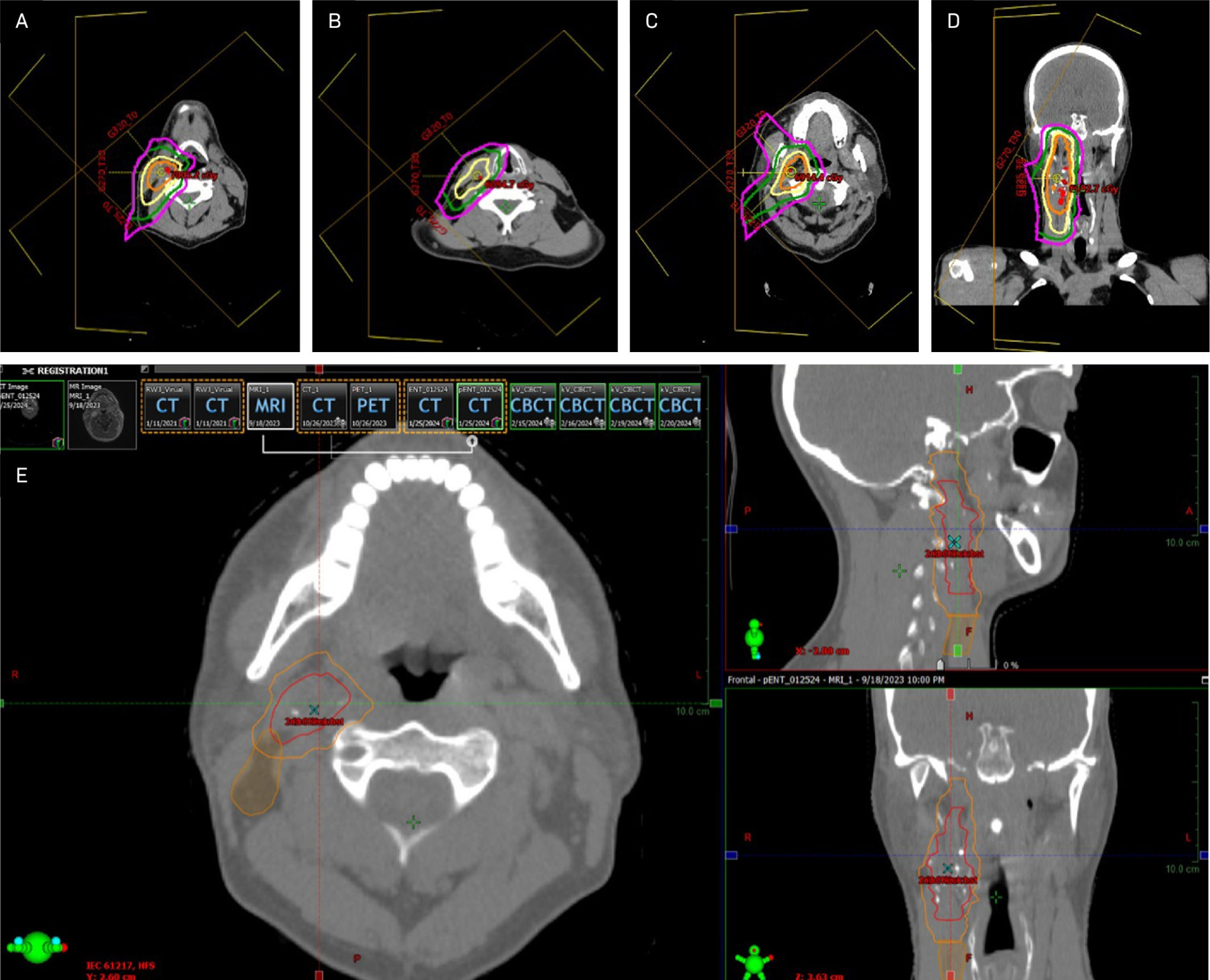
Complete surgical resection is the mainstay for curative treatment of MMNSTs, but the role and optimal approach of radiation therapy remain poorly defined due to the rarity of the tumors. For this patient, adjuvant radiation therapy was recommended due to the tumor’s locally aggressive behavior. Given the patient’s young age and ipsilateral treatment, intensity-modulated proton therapy was selected to maximize tumor coverage while sparing nearby and contralateral healthy tissues.
The most common types of radiation therapy (including conventional therapy, intensity-modulated radiation therapy, volumetric modulated arc therapy, and stereotactic body radiation therapy) use photon beams. As photons travel through tissue, they deposit energy gradually along their entire path, resulting in an exit dose, or radiation absorbed by healthy tissue on the path beyond the tumor.
Proton therapy achieves its tissue-sparing effect through a physical phenomenon called the Bragg peak. As charged particles, protons interact with electrons and nuclei while traveling through tissue, losing energy gradually at first. As they continue to slow down, these interactions increase, and eventually, the protons deposit the majority of their energy in a sharp peak (the Bragg peak) before stopping. This allows less radiation to pass beyond the tumor, sparing tissues on the contralateral side in this case. As part of radiation therapy, proton beam energy is precisely adjusted to position Bragg peaks at its target.
References
- Ship H, Shehadeh S, Montoya C. Malignant Melanotic Nerve Sheath Tumor of the Neck: A Case Report. Appl Radiat Oncol. 2025;14(2):1-7. doi:10.37549/ARO-D-25-00007.
Citation
. Virtual Rounds: Malignant Melanotic Nerve Sheath Tumor of the Neck 1. ARO Student Scan. 2026;3(3). doi:10.37549/VirtualRounds_Q4_2025.